The following protocol has been developed and optimized by R&D Systems IHC/ICC and stem cell laboratories for ICC experiments using stem cells grown on glass coverslips coated with stem cell subtype-specific substrates.
This protocol provides a basic guide for the preparation, fixation, and fluorescent staining of stem cells on glass coverslips. Each investigator must determine the precise experimental conditions required to generate a strong and specific signal for each antigen of interest. If R&D Systems primary antibodies are employed, please refer to the product datasheets to obtain the recommended working dilutions. In the staining protocol, signal visualization is achieved using R&D Systems NorthernLights™ range of fluorescent secondary antibodies and reagents. For all other reagents, please follow the manufacturer’s instructions.
Protocol for Coverslip Preparation Using Stem Cell Subtype-specific Substrates
Many cultured cell types do not adhere well to glass coverslips. The adhesion of many subtypes of cultured stem cells to glass is enhanced by adding a thin layer of the substrate on which the stem cells are normally cultured to a coverslip. Choose your typical stem cell culture substrate or follow the relevant procedure below for coating coverslips.
-
For use with embryonic stem (ES) or induced pluripotent stem (iPS) cells
-
For use with embryonic stem (ES) cells, induced pluripotent stem (iPS) cells, mesenchymal stem/stromal cells (MSCs), or neural stem cells (NSCs)
-
For use with mesenchymal stem/stromal cells (MSCs) cultured under serum-free conditions
-
For use with neural stem cells (NSCs)
Coverslip Coating Reagents Required for ES or iPS Cells Cultured on iMEFs
Reagents
-
Irradiated Mouse Embryonic Fibroblasts (iMEFs; Catalog # PSC001)
-
High glucose DMEM
-
L-Glutamine (200 mM)
-
Fetal bovine serum
-
Penicillin/Streptomycin (100X)
-
0.1% w/v solution of gelatin in sterile deionized H2O
-
95% EtOH
Materials
-
0.2 µm sterile filter unit
-
Coverslips (sterilized; Carolina Biological, Catalog # 633009 or equivalent)
-
Cell culture plate (24-well)
-
15 mL conical tubes
Equipment
-
37 °C, 5% CO2 incubator
-
37 °C water bath
-
Clinical centrifuge
Reagent Preparation
MEF Media - MEF media consists of high glucose DMEM, 10% fetal bovine serum, 2 mM L-glutamine, and if desired, a 1:100 dilution of penicillin/streptomycin (100X) stock. Filter sterilize the media using a 0.2 µm sterile filter unit.
Procedure for iMEF-coated Coverslip Preparation
-
Insert a sterile coverslip (sterilized with 95% EtOH and flamed) into each well of a 24-well plate.
-
Coat the appropriate number of plates for the desired number of cells by covering the surface of each well with 0.1% sterile gelatin for 15 minutes. For example, one vial of 6 x 10⁶ iMEFs can be plated on 2.5 24-well plates.
-
Warm the MEF media to 37 °C.
-
Thaw the desired number of vials of iMEFs by quickly warming the cryotube(s) in a 37 °C water bath until the cells are just thawed. Immediately transfer the contents of one vial to a 15 mL conical tube containing at least 5 mL of pre-warmed MEF media. Rinse the cryotube with an additional 1 mL of media to ensure the removal of all the cells.
-
Spin at 200 x g in a clinical centrifuge for 5 minutes.
-
Remove the supernatant and gently flick the pellet.
-
Aspirate the 0.1% gelatin from the plate(s).
-
Resuspend the iMEFs in MEF media and transfer to the gelatin-coated plates at a density of approximately 8 x 10⁴ cells/well of a 24-well plate.
-
Incubate overnight in a 37 °C, 5% CO2 incubator before seeding with stem cells.
-
Plate the cells at desired density.
View Fixation and Staining Procedures
Coverslip Coating Reagents Required for Stem Cells Cultured on a Defined Matrix
Reagents
-
StemXVivo™ Culture Matrix (Catalog # CCM013)
-
Sterile 1X PBS: 0.137 M NaCl, 0.05 M NaH2PO4, pH 7.4
-
95% EtOH
Materials
-
Coverslips (sterilized; Carolina Biological, Catalog # 633009 or equivalent)
-
Cell culture plate (24-well)
Equipment
Procedure for Defined Matrix-coated Coverslip Preparation
-
Insert a sterile coverslip (sterilized with 95% EtOH and flamed) into each well of a 24-well plate.
-
Thaw StemXVivo Culture Matrix at 2 °C to 8 °C before use.
-
Determine the volume of diluted culture matrix required. For example, approximately 400 µL for each well of a 24-well plate.
-
Dilute the Culture Matrix 1:100 in sterile 1X PBS. Mix gently. Do not vortex.
Note: The concentration of culture matrix used may need to be optimized for some cell types.
-
Immediately add diluted culture matrix to the plates. Gently sink the floating coverslips with a sterile pipette tip.
-
Incubate at 37 °C for 2-3 hours.
-
Immediately prior to plating the cells, remove the diluted culture matrix, and rinse once with sterile 1X PBS.
-
Plate the cells at desired density.
View Fixation and Staining Procedures
Coverslip Coating Reagents Required for MSCs Cultured under Serum-free Conditions
Note: MSCs sufficiently adhere to glass unless cultured under serum-free conditions (e.g., StemXVivo Serum-free Human MSC Expansion Media; Catalog # CCM014)
Reagents
-
Recombinant Human Fibronectin (Catalog # 4305-FN)
-
Sterile 1X PBS: 0.137 M NaCl, 0.05 M NaH2PO4, pH 7.4
-
95% EtOH
Materials
-
Coverslips (sterilized; Carolina Biological, Catalog # 633009 or equivalent)
-
Cell culture plate (24-well)
Equipment
Procedure for Fibronectin-coated Coverslip Preparation
-
Insert a sterile coverslip (sterilized with 95% EtOH and flamed) into each well of a 24-well plate.
-
Gently dilute the Fibronectin in PBS to a final concentration of 5 µg/mL.
-
Add 0.5 mL of the Fibronectin solution to each well. Gently sink the floating coverslips with a sterile pipette tip.
-
Incubate at room temperature for 3 hours or overnight at 2 °C to 8 °C.
-
Discard the Fibronectin solution and wash each well once with 1 mL of 1X PBS.
-
Plate the cells at desired density.
View Fixation and Staining Procedures
Coverslip Coating Reagents Required for NSCs
Reagents
-
Human Fibronectin (Catalog # 1918-FN-02M) or Bovine Fibronectin (Cataolog # 1030-FN)
-
Poly-L-ornithine
-
Sterile 1X PBS: 0.137 M NaCl, 0.05 M NaH2PO4, pH 7.4
-
95% EtOH
Materials
-
Coverslips (sterilized; Carolina Biological, Catalog # 633009 or equivalent)
-
Cell culture plate (24-well)
Equipment
Reagent Preparation
Poly-L-ornithine (1000X) - Dissolve Poly-L-ornithine in sterile PBS to make a 15 mg/mL stock. Aliquot and store at < -20 °C in a manual defrost freezer for up to 6 months. Avoid repeated freeze-thaw cycles.
Poly-L-ornithine Solution (1X) - Dilute 1000X Poly-L-ornithine 1000-fold in sterile PBS to make a 1X solution (15 μg/mL). Prepare fresh as needed.
Fibronectin Solution (1X) - Dilute the Human or Bovine Fibronectin in sterile PBS to make a 1 µg/mL solution. Mix by gentle swirling, without vortexing. Prepare fresh as needed.
Procedure for Poly-L-ornithine- and Fibronectin-coated Coverslip Preparation
-
Insert a sterile coverslip (sterilized with 95% EtOH and flamed) into each well of a 24-well plate.
-
Add 0.5 mL of 1X Poly-L-ornithine solution to each well. Gently sink the floating coverslips with a sterile pipette tip. Incubate overnight at 37 °C.
-
Discard the Poly-L-ornithine solution. Wash each well 3 times with 1 mL of sterile PBS each time.
-
Add 0.5 mL of sterile PBS to each well. Incubate overnight at 37 °C.
-
Discard the PBS. Wash each well once with 1 mL of sterile PBS.
-
Add 0.5 mL of 1 µg/mL Fibronectin solution to each well. Gently sink the floating coverslips with a sterile pipette tip.
-
Incubate in a 37 °C incubator for 3-24 hours.
-
Discard the 1X Fibronectin Solution and wash each well once with 1 mL of PBS.
-
Plate the cells at desired density.
Protocol for the Preparation & Fixation of Cells on Coverslips
Reagents Required
-
1X PBS: 0.137 M NaCl, 0.05 M NaH2PO4, pH 7.4
-
Formaldehyde Fixative Solution: 4% paraformaldehyde in PBS
-
Wash buffer: 0.1% BSA in 1X PBS
-
Cell culture medium
Procedure
-
When the cells have reached the desired density/age, remove the culture media from each well and wash twice with PBS.
-
Add 300-400 µL of 4% Formaldehyde Fixative Solution to each well, and incubate for 20 minutes at room temperature.
Note: Some cell types can be damaged by the change in surface tension that occurs when the culture medium is entirely removed and replaced with wash buffer. If this is the case, pre-fix the cells by adding 500 µL of 4% Formaldehyde Fixative Solution directly into the culture medium. After 2 minutes, replace the pre-fixation culture medium with 300-400 µL of 2% Formaldehyde Fixative Solution and incubate, for 20 minutes at room temperature.
-
Wash the wells twice with PBS and cover with 400 µL of wash buffer. The coverslips can be stored at 2 °C to 8 °C for up to 3 months or they may be stained immediately.
Note: Fixation can result in hydrophobic cross-linking of tissue proteins. The time, temperature, pH, and fixative used will determine the degree of cross-linking. Once the fixation protocol has been optimized, the same procedure should be used consistently.
Protocol for the Fluorescent ICC Staining of Cultured Cells on Coverslips
This protocol has been developed and optimized using R&D Systems NorthernLights fluorescent secondary antibodies but can be modified accordingly.
Reagents
-
Primary Antibodies
-
Blocking buffer: 10% normal donkey serum, 1% bovine serum albumin (BSA), 0.3% Triton® X-100 in 1X PBS
-
DAPI (4',6-diamidino-2-phenylindole) solution: Add 1 µL of 14.3 mM stock for every 5 mL of PBS. Store any unused DAPI at 2 °C to 8 °C, wrapped in aluminum foil
-
Deionized H2O
-
Dilution buffer: 1X PBS, 1% bovine serum albumin (BSA), 1% normal donkey serum, 0.3% Triton X-100, and 0.01% sodium azide
-
Anti-fade mounting medium
-
NorthernLights-conjugated secondary reagents (or equivalent)
-
1X PBS: 0.137 M NaCl, 0.05 M NaH2PO4, pH 7.4
-
Wash buffer: 0.1% BSA in 1X PBS
Materials
-
Cell-covered coverslips in a 6- or 24-well plate
-
Fine tweezers
Procedure
Note: This protocol is optimized for cells grown on coverslips in a 6- or 24-well plate but can be adapted accordingly.
-
Wash the coverslips containing the fixed cells 2 times in 400 µL of wash buffer.
-
Block non-specific staining by adding 400 µL of blocking buffer and incubate for 45 minutes at room temperature.
-
Remove blocking buffer. No rinsing is necessary.
-
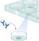
Dilute the unconjugated primary antibody (or fluorescence-conjugated primary) in dilution buffer according to the manufacturer’s instructions. For fluorescent ICC staining of cells on coverslips using R&D Systems antibodies, it is recommended to incubate at room temperature for 3 hours. Alternatively, incubate overnight at 2 °C to 8 °C.
Note: Appropriate controls are critical for the accurate interpretation of IHC/ICC results. All IHC/ICC experiments should include a negative control using the incubation buffer with no primary antibody to identify non-specific staining of the secondary reagents. Additional controls can be employed to support the specificity of staining generated by the primary antibody. These include absorption controls, isotype matched controls (for monoclonal primary antibodies), and tissue type controls.
-
Wash 2 times in 400 µL of wash buffer. If using a primary antibody with a direct fluorescent conjugate, go to step 8.
-
Dilute the secondary antibody in dilution buffer according to the manufacturer’s instructions. Add 400 µL to the wells, and incubate at room temperature for 1 hour in the dark. From this step forward samples should be protected from light.
Note: R&D Systems NorthernLights fluorescent secondary antibodies and streptavidin conjugates are bright, resistant to photobleaching, and are ideal for multi-color fluorescence microscopy.
Note: If a biotinylated antibody was used in step 4, apply the appropriate NorthernLights Streptavidin conjugate in step 6.
-
Rinse 2 times in 400 µL of wash buffer.
-
Add 300 µL of the diluted DAPI solution to each well, and incubate 2-5 minutes at room temperature. DAPI binds to DNA and is a convenient nuclear counterstain. It has an absorption maximum at 358 nm and fluoresces blue at an emission maximum of 461 nm.
Note: DAPI counterstain can obscure visualization of targets localized in cell nuclei.
-
Rinse once with PBS and once with water.
-
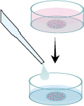
Carefully remove the coverslips from the wells and blot to remove any excess water. Dispense 1 drop of anti-fade mounting medium onto the microscope slide per coverslip. Mount the coverslip with the cells facing towards the microscope slide.
-
Visualize using a fluorescence microscope and filter sets appropriate for the label used. Slides can also be stored in a slide box at < -20 °C for later examination.
Note: Initial IHC/ICC studies often require further optimization and/or additionaltroubleshooting steps.
BG01V human embryonic stem cells are licensed from ViaCyte, Inc.
Triton is a registered trademark of Dow Chemical.